Welcome to week 26, and today we’re going to delve into stimulating muscle and from a neuromuscular assessment perspective. So what is muscle stimulation, how is it performed and what data can we derive from these assessments?
I spent a great deal of time during my PhD developing a new technique for the assessment of hamstrings neuromuscular performance via magnetic stimulation, so it’s a topic that I have a great interest in and one that I can hopefully start to unpack for you.
Transcutaneous Electrical Stimulation
I’m sure you’re well aware of muscle stimulation from the, somewhat debated, interferential treatment modality. Or indeed the most excellent (ahem!) Slendertone – other toning belts are available. The basic principle is that an electrical current is applied to the skin through an electrode and the underlying tissue, is ‘stimulated’. We’re not going to get into the debate of interferential or toning belts here; our focus is on the assessment of muscle function, which requires a rigorous and slightly different methodology.
Magnetic or Electrical Stimulation?
First off, there are a couple of ways in which we can evoke a contraction in a muscle or muscle group. We can use magnetic stimulation or electrical simulation. The inherent advantage of either technique over voluntary muscle activation is that they take the individual out of the equation i.e. you’re measuring a person’s physiologic capacity without the intrusion of conscious or unconscious inhibition.
Magnetic stimulation has the advantage that it is (by comparison to electrical) painless and you don’t need direct contact with the skin. A rapidly changing magnetic field within an encased copper coil when placed over a peripheral nerve causes its depolarisation and a twitch in the muscle/muscle group that it innervates [watch the clip below]. You may have heard of transcranial magnetic stimulation where the coil is placed over a specific area of the brain, with a variety of applications…?
Here we’re assessing quadriceps function by stimulating the femoral nerve. You can see we’ve also placed recording electrodes over the rectus femoris and vaustus laterals too (more on that later).
Electrical stimulation (ES) can elicit muscle activation either by stimulation of the peripheral nerve by a probe, or by placing electrodes over specific points over a the muscle itself. Electrical stimulation can be much more uncomfortable than magnetic since the electrical impulse is attenuated by the underlying tissue and thus to innervate deeper tissues a greater stimulation intensities are required. Sensory fibre endings are innervated and as such, it can be pretty painful. That said twitch responses are pretty well tolerated and ES is far cheaper than magnetic stimulation (MS).
Regardless of the method (MS/ES), the assessor must follow a systematic procedure to ensure that they locate the optimal site to stimulate the peripheral nerve and that the stimulation intensity is sufficient enough to produce a maximal (twitch) response.
Data Acquired From Muscle Stimulation
We can gain a lot of useful information about muscle function from evoked responses, particularly if we collect EMG (electromyographic) data too. Because of the technical difficulties (MS) or discomfort (ES) of inducing tetanic muscle contraction, we typically assess muscle twitch responses. These are elicited from either a single, 2 (doublet) or up to 8 (octet) supra-maximal stimulations just milliseconds apart.
Here’s a data trace of a quadriceps muscle twitch resulting from the stimulation of the femoral nerve in the inguinal region. You can see here that we’re able to assess:
- peak twitch force – how much force is produced,
- rate of force development – the speed of force production
- electromechanical delay – the ‘switch on time’ of the muscle
- Amplitude of the EMG response

We can also assess the relaxation parameters – how long it takes the force to dissipate, the ‘switch off time’ of the muscle, and the contraction time of the twitch (not shown).
What Does Information From Muscle Stimulation Mean?
We can use muscle stimulation techniques to assess numerous things. For example, the size, duration and relaxation of the twitch force can give us information about fatigue and fibre twitch properties. Smaller forces, longer contraction and relaxation times can indicate fatigue-related changes, and each of these indices infers something about where in the neuromuscular system that change may have occured.
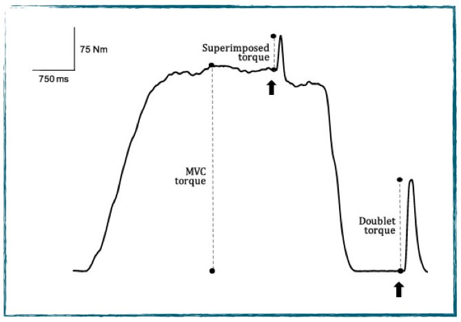
We can also stimulate a nerve during a maximal voluntary contraction (MVC) to add an additional action potential to those that are being produced voluntarily. This is called ‘superimposition’ or the interpolated twitch technique. Motor units that are not recruited voluntarily are stimulated and the added action potential may evoke an increase in force. This can give us information about inhibition – whether or not a person is able to recruit all of the available motor units, or if their maximal voluntary force production is inhibited.
Here’s an open access paper that investigated experimentally-induced anterior knee pain on voluntary activation as assessed by electrical stimulation (muscle not nerve).
The electromechanical delay (EMD) information from a single twitch can be used to assess the switch on time capacity of the musculature. If you want a quick reminder of what EMD is, see the post I wrote about it here. It’s really interesting to compare voluntary and evoked responses here. Typically we see that the EMD during evoked contractions is about half of that achieved under voluntary conditions, even though it’s only from a twitch! We first published data on this from our lab way back in 2007. There’s a link to that paper here if you’re interested to read it.
For example, voluntary EMD in recreationally-active individuals may be around 50 millisecond (ms) for the hamstrings, when evoked, this can be 25-30 ms. Likewise for quadriceps this could 25-30 ms and 10-15 ms for volitional and evoked, respectively.
Despite this voluntary / evoked differential, it is possible to decrease it and bring voluntary EMD closer to that of evoked. This requires specific training, and the result may prove useful in injury avoidance, especially when we consider the rapid time-frame over which injuries often happen.
I often refer to the responses under stimulated conditions as representing a potential ’emergency capacity’ that we’re perhaps not tapping in to under routine laboratory conditions. … Ever heard of the of stories where a mother runs out into the road to save her child – she lifts the car up with one hand and pulls the kid out with the other…? Well, maybe it’s not quite that dramatic, but nonetheless it’s interesting to know that we could have more in the tank than we realise.
Check out the Strength & Conditioning Course Listings